Digital movie acquisition allows precise quantification of different aspects of cell recruitment, such as speed of rolling or duration of arrest and microenvironmental positioning, by means of frame-by-frame automatic image computational analysis.
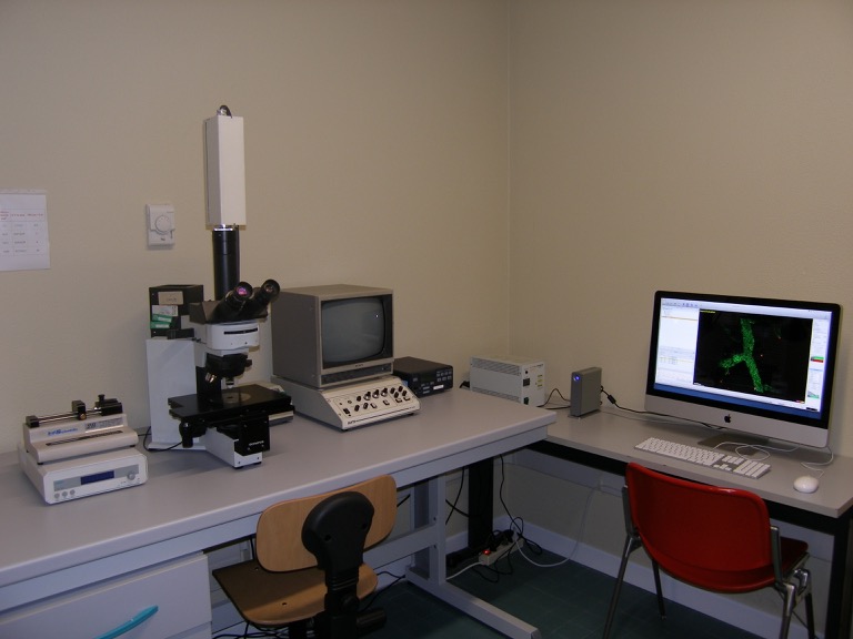
The intravital microscopy facility is complemented by the 2-photon microscopy facility, to study microenvironmental positioning of migrated cells.
Laudanna C, Constantin G. New models of intravital microscopy for analysis of chemokine receptor-mediated leukocyte vascular recognition. J Immunol Methods. 2003 Feb;273(1-2):115-23.
See intravital microscopy Movies
Administrated by prof. Gabriela Constantin’s Lab.
Hosted by: